
Page 68 - PRECONGRESS COURSE 04
P. 68
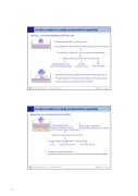
In-vitro models to study endometrial receptivity
Simon et al., J Clin Endocrinol Metab 82: 2607–2616, 1997
IL‐1a, IL‐1b
IL‐1ra 3
Embryos obtained after in vitro fertilization
EEC isolated from endometrium of fertile women (grown to confluence)
Co‐culture for 5 days (day 2 ‐ day 6 of embryonic development) analysis of EEC monolayers after embryo transfer
EEC with embryos that EEC with EEC reached the blastocyst stage arrested embryos without embryos
• The human blastocyst up‐regulates endometrial epithelial 3 subunit
• This up‐regulation is functionally relevant because it increases ability
of mouse blastocysts to adhere to EEC monolayer
University Hospital Essen - Prof. Dr. Ruth Grümmer ESHRE 2017 - Geneva
In-vitro models to study endometrial receptivity
Boggavarapu et al., Contraception 94:143–151 (2016)
Primary epithelial cells EEC and ESC isolated from
Basement membrane extract endometrium of fertile women (LH+4) Primary stromal cells in collagen gel
culture for 5 days culture with E2 and P4
+ thawed embryos obtained after in vitro fertilization
control + mifepristone (0.05 μM) + mifepristone (0.5 μM)
Analysis of embryo attachment
Analysis of endometrial cells for 16 reported endometrial receptivity markers
University Hospital Essen - Prof. Dr. Ruth Grümmer ESHRE 2017 - Geneva
68
